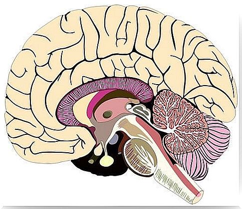
The Meninges: Structure And Functions
The brain and spinal cord are surrounded by three membrane-containing layers: the meninges. The three meninges are dura mater, arachnoid mater and pia mater. The last two consist of the lepto-meninges. Dura mater is also called the pachymeninx.
The main function of the meninges is to protect the brain. This is a very vulnerable organ that needs special protection. No other body needs it, at least not in the same way. In addition, these protective layers are part of the blood-brain barrier.
The meninges originate from another layer known as the primitive meninx, which consists of elements derived from the mesenchyme and neural crest. The primitive meninx is divided into two different layers: the inner (endomeninx) and the outer (ectomeninx).
Endomeninx is different from arachnoid mater and pia mater, and it comes from both the mesoderm and the ectoderm. On the other hand, the ectomeninx forms the dura mater and the bones of the neurotransmitter, and consists only of the mesoderm.
Structure of the meninges
Dura mater
The dura mater is the outer layer of the meninges and has two layers. The outer layer is the periosteum and contains blood vessels and nerves. It adheres to the inner surface of the skull, with joints adapted specifically to the skull.
The name of the deepest layer of dura mater is the meningeal layer. It is responsible for the reflexes that divide the brain into space.
Among these rooms are the most prominent: falx cerebri and tentorium cerebelli. In addition, there is no distinct boundary between the dura mater and the periosteum. The layers differ histologically from the fact that the meningeal layer has fewer fibroblasts and relatively less collagen (2).
Arachnoid mater
Arachnoid feeder is the middle layer of the meninges. It contains subarachnoid space which in turn stores cerebrospinal fluid (CSF). The depth of subarachnoid space varies depending on the relationship between the arachnoid feeder and the pia feeder.
Two separate cell layers form the arachnoid mater. The arachnoid barrier cell layer lies along the edge of the cells in the dura mater (3). This layer is full of cells that are closely linked by many desmosomes and tight junctions. Thus, this layer prevents liquid from moving through it.
The reticular arachnoid layer is deep inside the arachnoid feeder. Its cells connect the subarachnoid space and attach to the pia mater. They also follow the blood vessels that run through the layer (1).
Arachnoid villi are microscopic structures that play an important role in the absorption of CSF. However, the way they work is unclear. Some believe that arachnoid villi may also play a role in regulating CSF volume.
The third of the meninges: Pia feeds
Pia mater is the innermost layer of the meninges. It is a delicate vascular structure that surrounds and attaches to the surface of the brain and spinal cord and protects it.
It creates a continuous layer of cells that attaches to the surface of the brain and then sinks into the cracks and grooves in the brain. Desmosomes and gap connections bind the cells, which means that the layer can act as a barrier.
The Virchow-Robin space
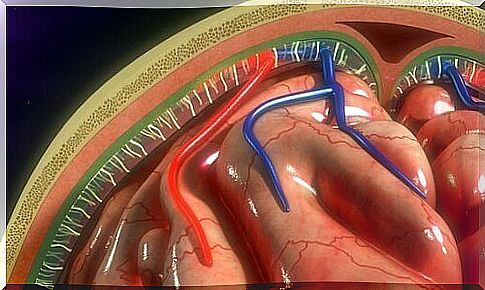
The Virchow-Robin space is located around the blood vessels and surrounds the small arteries and arterioles. They drill through the surface of the brain and extend inward from the subarachnoid space (1).
These gaps increase in size as they age, without an apparent associated loss of cognitive function (4). In addition, the dilation of these chambers is related to conditions such as hypertension, neuropsychiatric disorders, multiple sclerosis and trauma (5).
Finally, the authors Patel and Kirmi (2009) emphasized the importance of knowing the meninges. It is important to understand their structure, functions and anatomy because it will help us understand the pathology associated with the meninges.