Biopsychological Research Methods: What Are They Really?
Biopsychological research methods have evolved in recent decades. Although there are several of them, in this article we will focus on those who study what happens in the brain under certain conditions.
Authors such as Dewsbury (1991) define biopsychology as “the scientific study of behavioral biology “, a field also called psychobiology. However, other authors prefer the term biopsychology because it “indicates a more biological approach to the study of psychology, than a psychological approach to the study of biology.”
The stimulation and visualization methods of the human brain
Observation and recording of brain activity is a very important milestone that was achieved thanks to the various techniques that scientists developed during the 20th century. Biopsychological research methods are without a doubt a breakthrough in the study of our most curious body.
Biopsychological research methods: Contrast radiography
This technique consists of injecting a substance into the body to absorb X-rays. In this way, the researchers see the contrast between the different rooms and the surrounding tissue.
Cerebral angiography is a type of contrast radiography. To do this, a contrast medium is inserted into a brain duct. The goal is to observe the circulatory system while performing X-rays. This technique is very useful for locating ductal lesions and brain tumors.
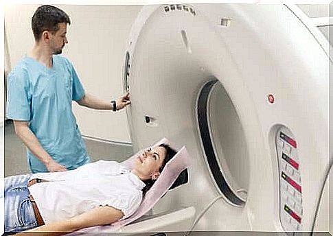
Computerized axial tomography scan (CT scan)
Through a CT scan, experts can see the entire brain structure. During the test, the patient lies in the middle of a large cylinder. While the patient is lying still, an X-ray tube and a receptor take many separate photographs. This happens while the emitter and receptor spin around the patient’s head.
All this information is transferred to a computer, which allows doctors to explore the brain on a horizontal plane. They usually do this on eight to nine horizontal brain sections. When all the discoveries are combined, it is possible to create a three-dimensional representation of the brain.
Nuclear Magnetic Resonance (NMR)
NMR enables high-resolution image processing thanks to the different waves emitted by hydrogen atoms when activated by the radio frequency on a magnetic field. It provides a high spatial resolution and produces three-dimensional images.
Biopsychological research methods: positron emission tomography (PET)
In biopsychological research methods, PET provides images of brain activity instead of brain structure. To get the images, the researchers inject radioactive fludeoxyglucose (FDG) into the carotid artery. Active neurons rapidly absorb FDG, which accumulates when the neurons no longer metabolize it. Then it decomposes slowly. This way you can observe which neurons are active at a given time during different activities.
Functional magnetic resonance imaging (fMRI)
On the other hand, MRI provides a picture of the increase in the amount of oxygen present in the blood of the brain. Thus, it measures brain activity in a successful way. If we compare it to PET, it actually has four advantages:
- Doctors do not need to inject the patient with anything.
- It provides both functional and structural information.
- It provides a better spatial resolution.
- Provides three-dimensional images of the entire brain.
Biopsychological research methods: magnetoencephalography (MEG)
It measures the changes in the magnetic fields on the scalp. These changes occur due to the variations in the guidelines of neuronal activity.
Transcranial magnetic stimulation (TMS)
Walsh and Rothwell (2000) state that TMS “changes an area of the cortex, creating a magnetic field under the coil that passes over the skull.” TMS “turns off” a part of the brain temporarily, to study behavior and cognition under these circumstances.
Lesion methods
Lesion methods focus on the destruction of a small area of the brain to see how it affects behavior.
- Aspiration lesion. This method creates a lesion in an exposed or easily accessible cortical tissue area. Doctors remove the tissue with a crystal pipette.
- Radio frequency damage. It is performed by making small subcortical lesions. For that, an electrode can channel the high frequency current through the tissue of interest. The size and shape of the lesion depends on three factors:
- Duration of the procedure.
- Intensity of the day.
- Configuration of the electrode point.
- Scalpel cut. It consists of slicing the brain area of interest.
- Cooling damage. This regulatory method for biopsychology, although included under lesion methods, is in fact temporary and reversible. Instead of destroying tissue, an area is cooled slightly above freezing. Neurons stop sending signals, so the cold brain area remains blocked. With this, researchers can see what behavioral changes are caused by these areas. When the temperature returns to normal, brain function is restored .
Biopsychological research methods: Electrical stimulation
Another biopsychology and regulatory method is electrical stimulation. The procedure consists of electrical stimulation of a nervous system structure to obtain data on its functions. Usually a bipolar electrode is used.
This stimulation “shoots” neurons and changes their behavior. In general, it tends to have the opposite effect of the lesion method. For example, if it is possible to drastically reduce sleep hours with a lesion, the sleep pattern may become uncomfortable and more difficult to control with electrical stimulation.
Lesion methods with electrical recording
- Intracellular recording. This technique is performed by inserting a microelectrode into the interior of a neuron. It registers fluctuations of membrane potential.
- Extracellular device recording. A microelectrode is placed in the extracellular fluid that surrounds the neuron. It does not provide information on membrane potential.
- Multimedia recording. In this case, the electrode point is larger than a microelectrode, so that it captures signals from many neurons simultaneously. The discovered potentials then go on a circuit that integrates them.
- Invasive EEG monitoring. The stainless steel electrodes enter the skull. For subcortical signals, the usual electrodes are made of cable and implanted through stereotactic surgery.
Biopsychological research methods: A long way to go
In this article, we talked about the most important things in biopsychological research methods. However, it is also worth mentioning that there are other biopsychological research methods that study other areas of the body, such as muscle tension measurements, eye movement recordings, skin conductivity or cardiovascular activity.
The breakthrough in this field has undoubtedly been spectacular, but not decisive. Maybe in a few years, researchers will come up with new techniques that will contribute to the development of neuroscience. It will help improve the quality of life of many people who are affected by neural changes.